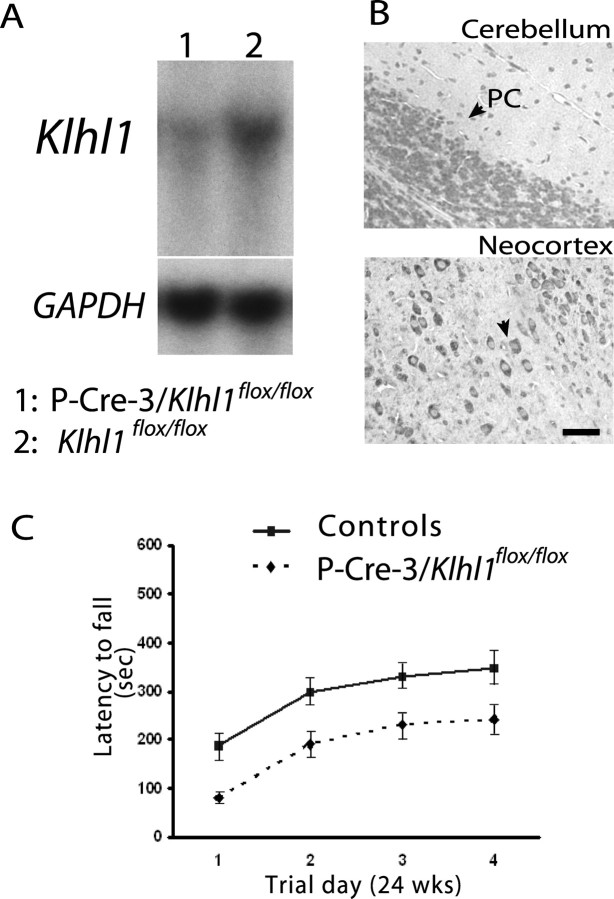
Figure 4.
Analyses of mice in which Klhl1 is deleted only in cerebellar Purkinje cells. A, Northern blot analysis demonstrated that the mRNA Klhl1 is dramatically decreased in cerebellar tissue from 24-week-old homozygous P-CRE-3/Klhl1flox/flox mice (lane 1) compared with the controls (Klhl1flox/flox; lane 2). The RNA loading levels of each lane were measured by a subsequent hybridization with GAPDH probe. B, Paraffin-embedded sections of brain from homozygous P-CRE-3/Klhl1flox/flox mice stained for Klhl1 using 13D9 mAb show cerebellar Purkinje cell-specific Klhl1 deficits. There was an absence of staining of Klhl1 in the soma and dendrites of Purkinje cells (left), but Klhl1 is normally expressed in the other neurons in this brain (neocortex; right). PC, Purkinje cell. Scale bar, 25 μm. C, Motor coordination performance deficits of 24-week-old P-CRE-3/Klhl1flox/flox mice on accelerating rotarods. Their performance was significantly worse (p = 0.011 at day 4) than the performance of controls. Error bars indicate SEM.

