
Figure 3.
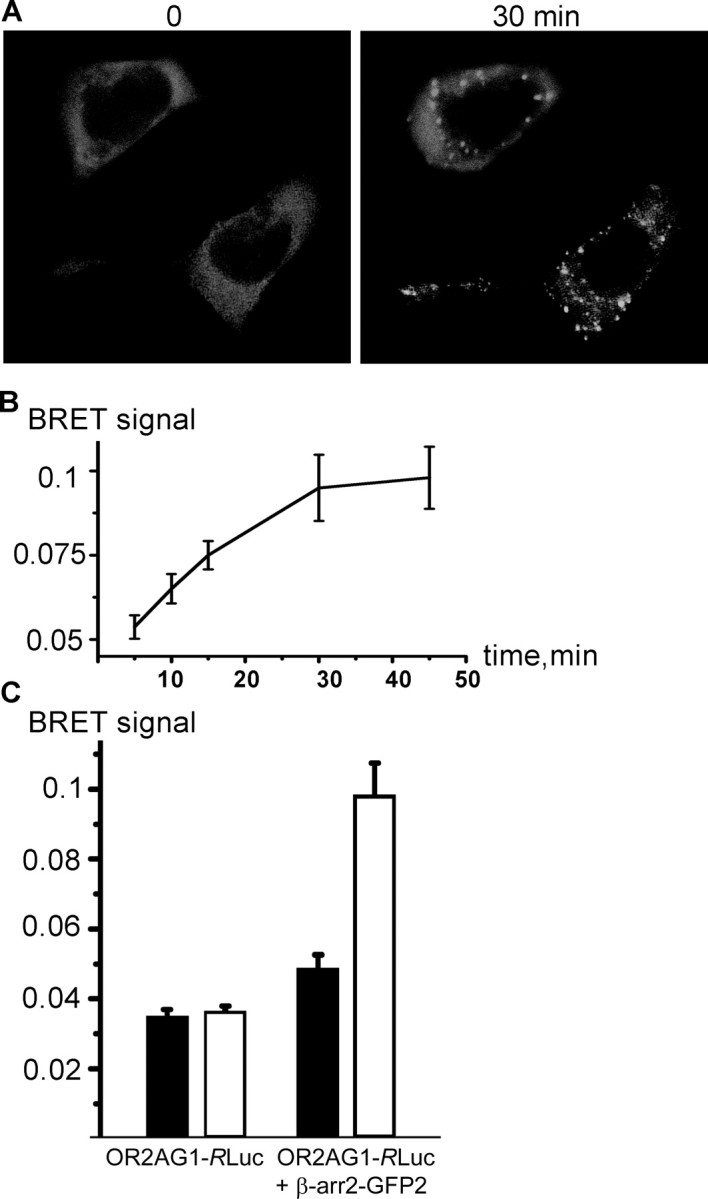
β-arrestin2 interacts with olfactory receptors during ligand binding. A, Confocal microscopy of HEK293 cells cotransfected with OR2AG1 and β-arrestin2–GFP. Amylbutyrat treatment (30 min) causes translocation of β-arrestin2 into intracellular vesicular structures. B, Cells were cotransfected with β-arrestin–GFP and OR2AG1–RLuc fusion constructs, and the BRET ratio was measured at different time points of amylbutyrat stimulation 48 h after transfection. The BRET signal increased in a time-dependent manner during addition of the agonist. Data are the mean from five independent experiments. C, Cells were cotransfected with β-arrestin–GFP and OR2AG1–RLuc fusion constructs or with OR2AG1–RLuc alone. The BRET ratio was determined 48 h after transfection on amylbutyrat-stimulated (white bars) and nonstimulated (black bars) cells. No significant change of the BRET ratio during addition of amylbutyrat is observed in cells expressing OR2AG1–RLuc alone. In contrast, addition of the agonist to cells expressing the OR2AG1–RLuc fusion construct together with β-arrestin–GFP leads to an increase of the BRET signal. Error bars represent SEM.