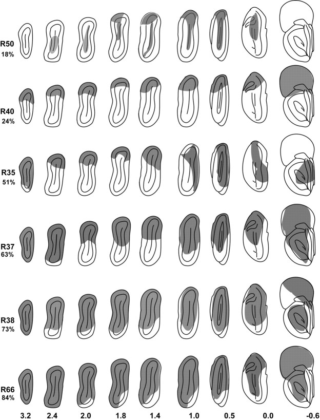
Figure 5.
Diagrammatic representations of frontal sections of the olfactory bulb for six experimental rats showing the areas removed by the lesion (shaded regions). The percentage of the mitral cell layer removed is given under the rat identification numbers on the left. Numbers on the bottom are millimeters from the rostral aspect of the accessory olfactory bulb. Dorsal is to the top, and medial is to the right in each section drawing.

