
Figure 1.
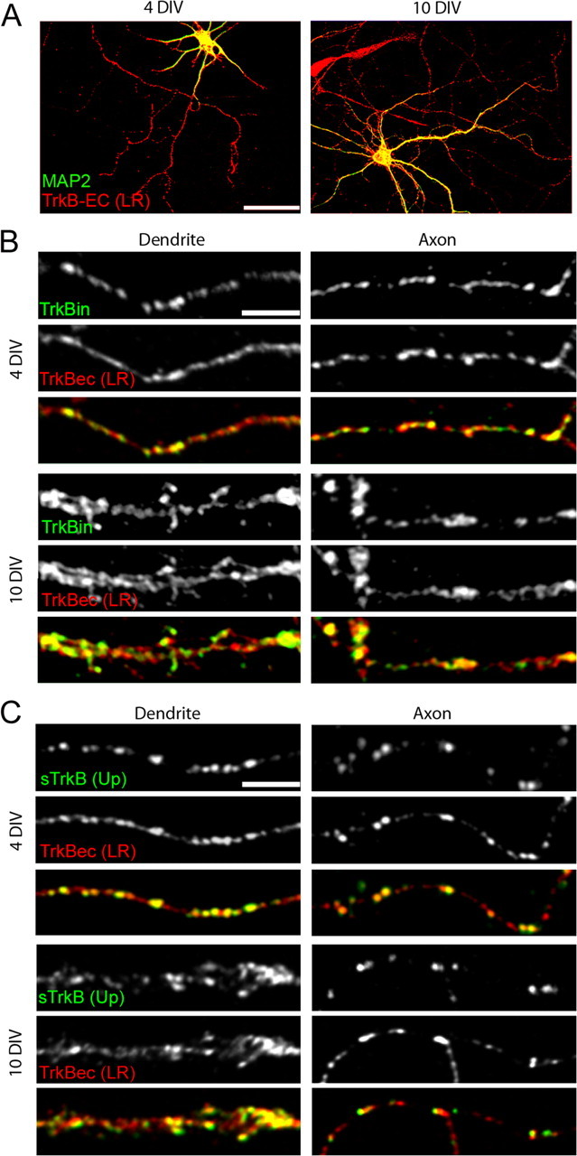
TrkB is found in both axons and dendrites of cortical neurons. A–C, Neurons were labeled with an antibody to the extracellular domain of TrkB [TrkBec (LR); A–C, red] and MAP2 (dendritic marker; A, green), full-length TrkB (TrkBin; B, green) or surface TrkB [sTrkB using TrkBec (Up); C, green]. Colocalization is indicated by yellow in the overlays. A, TrkB is present in axons, dendrites, and cell bodies before (4 DIV) and at the peak of synapse formation (10 DIV) in cortical cultures. B, Full-length TrkB (TrkBin) is expressed in both dendrites and axons and is highly colocalized with TrkBec (LR), suggesting that TrkBec is a reliable indicator of the presence of TrkBin in cortical neurons at the ages examined. In dendrites, TrkB shows a punctate pattern over a low diffuse background, and in axons, TrkB is mainly punctate at both 4 and 10 DIV. C, Surface TrkB [sTrkB; TrkBec (Up)] is widely distributed in puncta on both dendrites and axons and appears to be a subset of the total TrkB [labeled here with TrkBec (LR)]. Scale bars: A, 50 μm; B, C, 5 μm.