Figure 2.
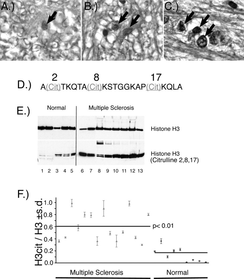
Citrullinated histones in normal and MS brain and nuclear fractions. Immunohistochemical staining of normal (A, 400×), and MS (B, 400×; C, 1000× oil) white matter with anti-citrulline (F95) monoclonal antibody. Arrows indicate nuclear labeling with the F95 antibody. A more intense nuclear labeling was observed in the MS white matter. D, The sequence of the tri-citrullinated N-terminal histone H3 peptide in which the arginyl residues at positions 2, 8, and 17 are replaced by citrulline. E, Western immunoblot of nuclear fractions prepared from white matter from normal and MS individuals with anti-histone H3 and anti-H3cit. F, The ratio ± SD of H3cit/H3 in nuclear white matter fractions from normal and individuals with MS. The mean of the H3cit/H3 ratios for the MS and normal groups is indicated by the horizontal lines (p < 0.01, nonparametric test). A list of the clinical diagnosis for each of the samples used in these experiments and the values ± SD of the H3cit/H3 ratios are provided in supplemental Table 1 (available at www.jneurosci.org as supplemental material).

