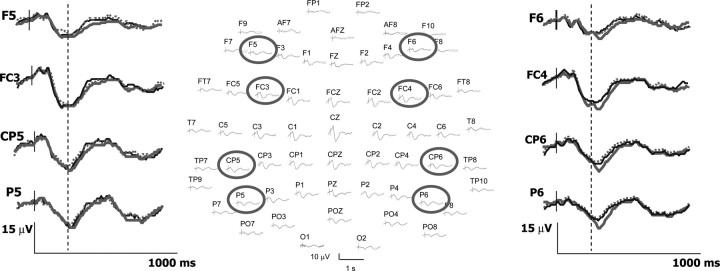
Figure 5.
Cortical responses to painful stimuli associated or not to emotional modulation of pain. The 64-channel SEPs at their scalp positions (nose up) are represented in the center. Positive voltages are represented downward. Traces from eight selected electrodes (circles) are enlarged in the left and in the right parts to illustrate that SEP amplitudes to painful stimuli were significantly enhanced during the unpleasant-body condition (gray traces) relative to both unpleasant-nonbody (dotted) and pleasant-body (black) conditions. Note also that amplitude differences occurred only after 270 ms poststimulus (dotted line) and concerned exclusively the electrodes located on the right side of the scalp. See Results for details.

