
Figure 2.
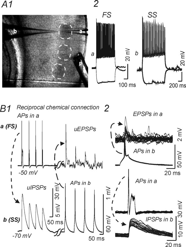
Recordings form reciprocally connected spiny stellate cell and fast-spiking interneuron in layer IV barrel. A1, Low-magnification image of the barrel field in a thalamocortical slice, with pial surface to the right. Dashed circles indicate individual barrels. a, b, Recording patch pipettes within a barrel near the layer V border. P, Local perfusion electrode. A2, B, Current-clamp recordings. A2, Responses evoked by current steps (−100 and +200 pA) in fast-spiking interneuron (a) and regular-spiking spiny stellate cell (b). B, Paired recordings from a reciprocally connected fast-spiking cell (a) (FS) and spiny stellate cell (b) (SS) show unitary synaptic potentials elicited by trains (B1) or single action potentials (B2) in the presynaptic cell. B1, Trains of APs elicited by depolarizing currents (250 pA; 100 ms) in cell a (top left) evoked uIPSPs in cell b (bottom left, dotted line and arrow), whereas action potentials in cell b (bottom right, dashed line and arrow) elicited uEPSPs in cell a (top left). B2, Top, Single APs in cell b evoked uEPSPs in cell a (top panel, 15 traces overlaid). B2, Bottom, Single APs in cell a evoked uIPSPs in cell b (15 traces overlaid). Calibrations in bottom right panel for traces showing APs; calibrations in bottom left panel for traces showing uPSPs.