
Figure 6.
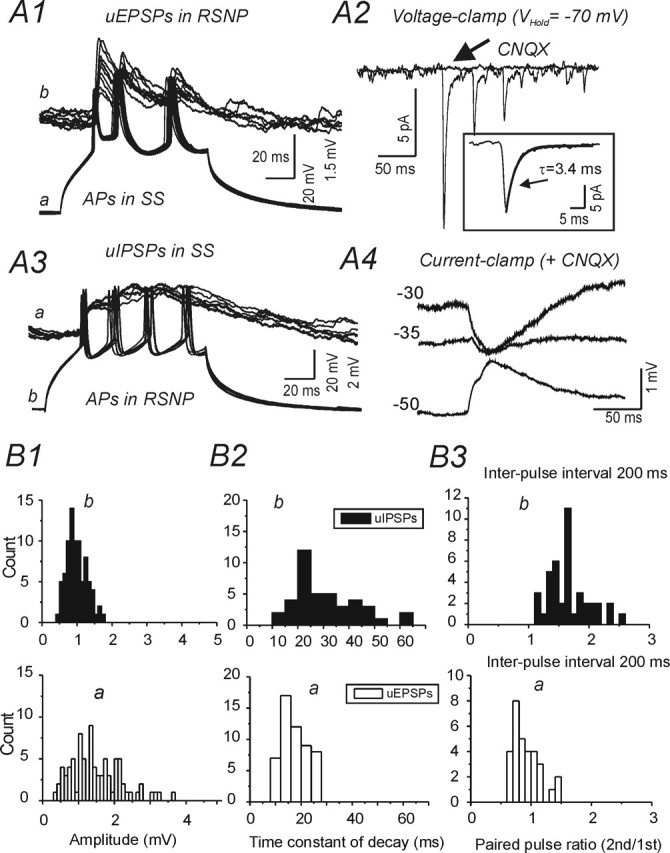
Reciprocal connections between spiny neurons and regular spiking (RSNP) interneurons. A1, Depolarizing current pulses (300 pA; 150 ms) evoke trains of three APs in SS cell a (bottom) and trains of three uEPSPs in RSNP cell b (top panel, 30 traces superimposed). A2, Voltage-clamp recording of uEPSCs in RSNP cell b evoked as in A3 under control conditions, and in the presence of CNQX, which abolished the uEPSCs (arrow). Inset, Averaged initial uEPSC of 30 trains with overlaid single exponential fit for decay phase (solid gray line; τ = 3.4 ms; Vhold = −70 mV). A3, Depolarizing current pulses (300 pA; 150 ms) evoke trains of four APs in RSNP cell a (bottom) and depolarizing uIPSPs in RSNP cell b (top). A4, Current-clamp recording of uIPSPs at indicated Vm values in the presence of CNQX. B, Histograms showing distributions of amplitude (B1), decay time constant (τD) (B2), and paired-pulse ratio (at 200 ms interval) (B3) for uEPSPs in RSNP cell a of A1 (open bars, top graphs) and uIPSPs in an SS cell a of A3 (black bars, bottom graphs). ECl = −32 mV in A3 and A4