
Figure 1.
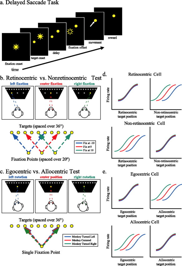
Tasks. a, Delayed saccade trials. Subjects fixated (±2°) a centrally located yellow LED. After a delay, a target LED was illuminated; subjects were required to maintain fixation until the central light was extinguished (200–600 ms) and then shift gaze to the target (2–3°) and maintain fixation of it for 500 ms to receive a juice reward. b, Retinocentric versus nonretinocentric test. The fixation LED appeared at either the center of the LED panel or 10° to the left or right of the center. Targets were drawn from an array of 10 LEDs, spaced 4° apart from −18 to 18° and typically 10° above or below the array of fixation points. c, Egocentric versus allocentric test. Subjects performed delayed saccades from the central fixation point to the array of 10 targets after passive whole-body rotation 20° in either direction around the vertical axis. d, Expected tuning curves in the retinocentric versus nonretinocentric test. Tuning curves, separated by fixation point, are expected to align when plotted as a function of retinocentric target position if these neurons encode only information about the saccade vector/retinal vector. If these neurons encode information with respect to the subject but not the eye, tuning curves will align when plotted as a function of the nonretinocentric target position. e, Expected tuning curves in the egocentric versus allocentric test. Tuning curves, separated by rotation, are expected to align when plotted as a function of target position with respect to the subject if these neurons encode spatial information in egocentric coordinates. If these neurons encode spatial information allocentrically, the tuning curves will align when plotted as a function of the target position in the room.