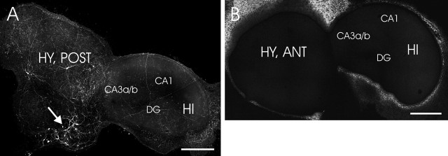
Figure 1.
Representative images of histamine immunostaining in a coculture of the hippocampus and hypothalamus. A, Histaminergic neurons (arrows), which are located in the posterior hypothalamic slice, distribute their neurites all over in HI plus HY (POST) (7 d in vitro). B, No histamine immunopositive staining was detected in HI plus HY (ANT). Different subregions of the hippocampus CA3a/b, CA1, and dentate gyrus are indicated. Scale bars, 500 μm. DG, Dentate gyrus.

