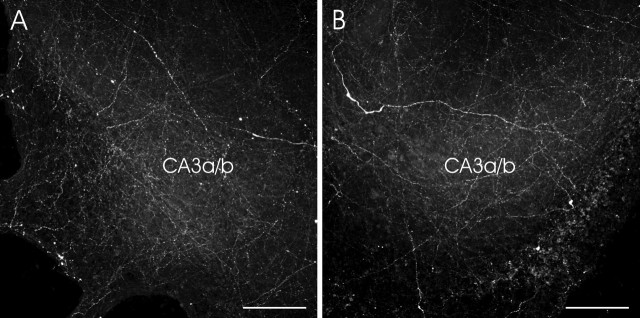
Figure 2.
Representative images showing the histaminergic innervation in the CA3a/b subregion of the hippocampus. A, In HI plus HY (POST) (9 d in vitro), histaminergic fibers grew to the hippocampal slice. B, HI plus HY (POST) (7 d in vitro) was treated with KA (5 μm) for 12 h and, after the medium change, further cultured for 48 h. Note that the density of fluorescent, histamine-containing fibers in the CA3a/b region did not differ in these different experimental conditions. Scale bars, 100 μm.

