Figure 3.

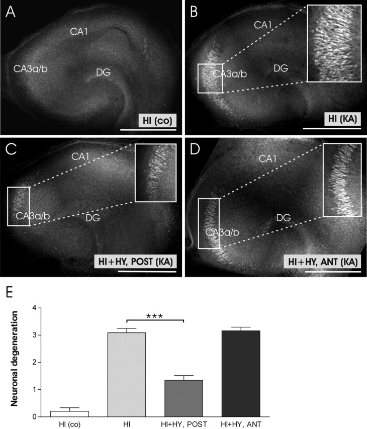
Representative confocal images of KA-induced neuronal degeneration in the hippocampal slices cultured for 7 d alone, or together with the posterior or anterior hypothalamic slice. A, A representative image of control HI (no KA treatment) shows no FJB-stained neurons. B, HI was treated with KA (5 μm) for 12 h. Note the extensive staining of pyramidal neurons in the CA3a/b region. C, HI plus HY (POST) (7 d in vitro) was treated with KA (5 μm) for 12 h. The area of FJB-stained, degenerating neurons in the Ca3a/b region is smaller when compared with Figure 3B. D, HI plus HY (ANT) (7 d in vitro) was treated with KA (5 μm) for 12 h. No clear decrease in neuronal damage is seen in the CA3a/b region when compared with Figure 3B. E, According to our scoring system, KA-induced neuronal damage was significantly decreased in the CA3a/b region of HI plus HY (POST), whereas no significant decrease was detected in HI plus HY (ANT) compared with KA-treated HI. The results are given as means ± SEM. co, Control (not KA treated); DG, dentate gyrus. ***Significant difference of p < 0.001 (one-way ANOVA). Scale bars, 500 μm.
