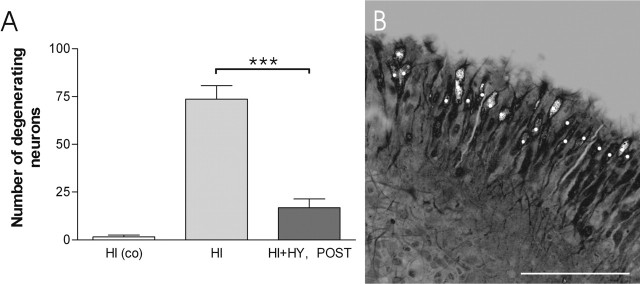
Figure 4.
The number of FJB-stained, degenerating neurons in the CA3a/b region after the KA treatment. A, HI and HI plus HY (POST) (7 d in vitro) were treated with KA (5 μm) for 12 h, and FJB-stained, degenerating neurons were counted in a 250 μm2 area of the CA3a/b region. A significant decrease was detected in the number of FJB-stained neurons in HI plus HY (POST) compared with HI. Control HI were cultured in normal culture medium and not treated with KA. ***Significant difference of p < 0.001 (one-way ANOVA). co, Control (not KA treated). B, A representative confocal image of a single 250 μm2 optical section of the CA3a/b region in HI (7 d in vitro), which was treated with KA for 12 h. Counted cells at this level are marked with the white dots. The results are given as means ± SEM. Scale bar, 100 μm.

