
Figure 5.
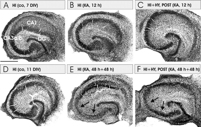
Representative images of thionin-stained cultures of HI, HI plus HY (POST), and HI plus HY (ANT). A, The control (co) HI cultured for 7 d in normal medium. B, HI was treated with KA (5 μm) for 12 h. No decrease in neuronal density was detected. C, HI plus HY (POST) treated with KA for 12 h. This figure corroborates that decrease in FJB-stained neurons in the coculture with histaminergic neurons (Figs. 3, 4) is not caused by the decrease in neuronal density in the CA3a/b area. D, The control HI cultured for 11 d in normal medium. E, HI was treated with KA (5 μm) for 48 h and followed by a 48 h recovery period in the normal culture medium. Note that pyramidal neurons have disappeared in the CA3a/b region (arrows). F, HI plus HY (POST) treated with KA (5 μm) for 48 h followed by a 48 h recovery period in the normal medium. CA3a/b and CA3c pyramidal neurons are clearly better preserved in F than in E (arrows). Scale bar: (in A) A–F, 500 μm.