
Figure 1.
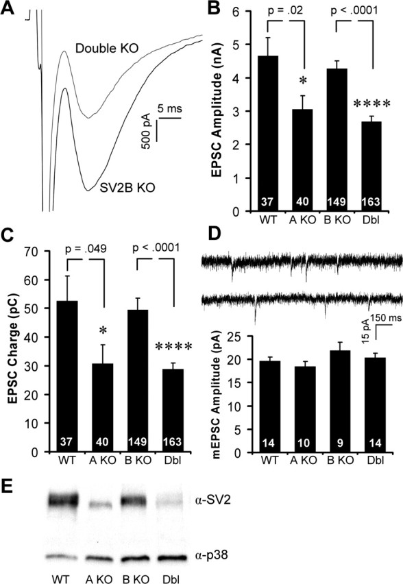
Excitatory responses are smaller in neurons lacking SV2A. Hippocampal neurons cultured from SV2 knock-out mice were analyzed in the whole-cell voltage-clamp configuration. Neurons were held at −60 mV, and single EPSCs were evoked by depolarizing for 1 ms. Graphs show means ± SEM. The number of cells analyzed is indicated within the bars. A, Sample traces from SV2B knock-out and SV2A/B double knock-out neurons. B, C, Average EPSC peak amplitudes (B) and total EPSC charge (C) for wild-type and SV2 knock-out neurons. Data are from littermate cultures of six WT and SV2A KO animals and 23 SV2B KO and double (Dbl) KO animals. D, Top, Representative traces of spontaneous mEPSCs recorded from SV2B and SV2A/B double knock-outs. Bottom, Mean amplitudes of mEPSCs were unchanged in wild-type and knock-out neurons. Data are from four WT, three SV2A KO, three SV2B KO, and three double KO cultures. E, Immunoblot analysis of cultured wild-type and SV2 knock-out cultures probed with anti-synaptophysin (p38) and anti-SV2 reveal that SV2A is the predominant isoform in these cultures.