
Figure 5.
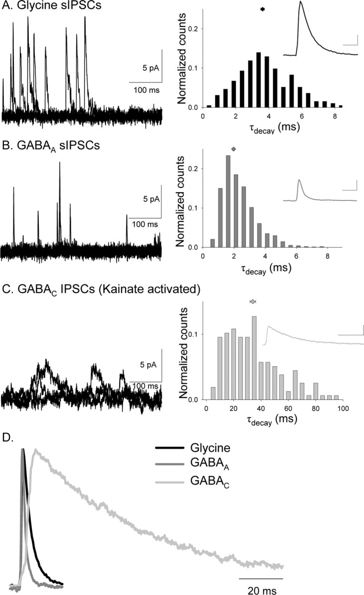
Glycine, GABAA, and GABAC receptors mediate sIPSCs with distinct kinetics in rod bipolar cells. A, Examples of isolated glycine receptor sIPSCs that were measured in the presence of bicuculline and TPMPA (left). The τdecay histogram distribution (normalized to the total number of events) for all glycine receptor-mediated sIPSCs recorded are shown in the right, and the inset shows the average glycine receptor-mediated sIPSC. Calibration:2 pA, 5 ms. B, Examples of isolated GABAA receptor sIPSCs that were measured in the presence of strychnine and TPMPA (left). The normalized τdecay histogram distribution for all GABAA receptor-mediated sIPSCs recorded are shown in the right, and the inset shows the average GABAA receptor-mediated sIPSC. Calibration: 2 pA, 5 ms. GABAA receptor-mediated sIPSCs had a significantly shorter τdecay than glycine receptor-mediated sIPSCs (K–S, p < 0.0001). C, Examples of isolated GABAC receptor sIPSCs, measured in the presence of kainate, strychnine, and bicuculline (left). The normalized τdecay histogram distribution for all GABAC receptor-mediated sIPSCs recorded is shown in the right, and the inset shows the average GABAC receptor-mediated sIPSC. Calibration: 2 pA, 50 ms. GABAC receptor-mediated sIPSCs had a significantly longer τdecay than glycine (K–S, p < 0.0001) and GABAA (K–S, p < 0.0001) receptor-mediated sIPSCs. D, Average normalized sIPSCs mediated by glycine, GABAA, and GABAC receptors. GABAC receptor-mediated sIPSCs have the longest decay time.