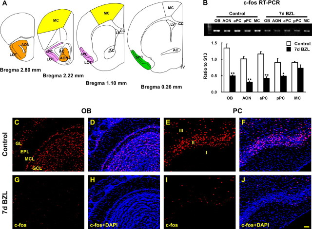
Figure 1.
Reduction of c-Fos protein and mRNA expression in OB and PC after bilateral zinc sulfate lesion. A, The diagram depicts the areas where we microdissected AON (orange), aPC (pink), pPC (green), and MC (yellow). B, The amount of c-fos mRNA was measured by semiquantitative RT-PCR and was reduced 7 d after bilateral zinc sulfate lesion (BZL) in OB, AON, aPC, and pPC but not in MC [two-way ANOVA across all tissues and lesions; F(4, 30) = 6.80; p < 0.01; post hoc Tukey’s comparisons revealed significant differences between control and BZL groups in these olfactory tissues (*p < 0.05; **p < 0.01)]. Data are means + SEM (n = 4). c-Fos immunostaining was visualized by confocal microscopy (red) and nuclei were counterstained with DAPI (blue). C–J, Seven days after BZL, c-Fos immunoreactivity was markedly reduced in OB (G, H) and PC (I, J) compared with the control OB (C, D) and PC (E, F). Scale bar, 50 μm. 3V, Third ventricle; AC, anterior commissure; CC, corpus callosum; EPL, external plexiform layer; GCL, granule cell layer; GL, glomerular layer; LOT, lateral olfactory tract; LV, lateral ventricle; MCL, mitral cell layer.

