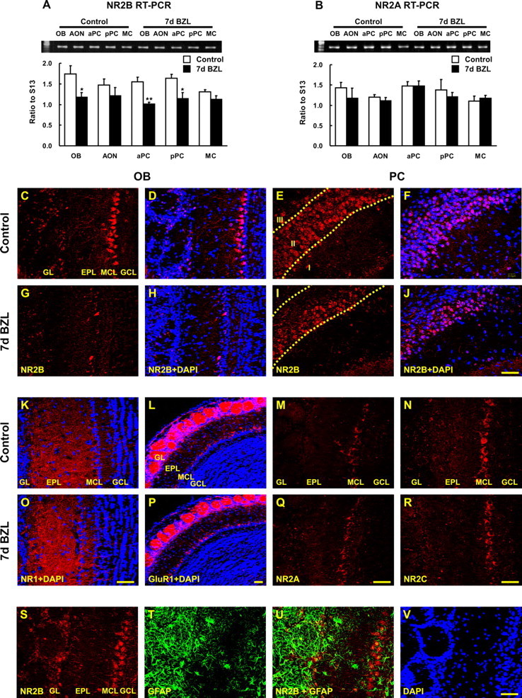
Figure 2.

Expression of glutamate receptors in OB and PC after bilateral zinc sulfate lesion. The amount of NR2B and NR2A mRNA expression was measured by semiquantitative RT-PCR. A, B, NR2B expression declined 7 d after bilateral zinc sulfate lesion (BZL) (A) in OB, aPC, and pPC [two way ANOVA across all tissues and lesions, F(4, 30) = 0.91, p = 0.47, no interaction between lesion and tissue; one-way ANOVA for the main effect of the lesion, F(1, 30) = 25.18, p < 0.01; post hoc Tukey’s comparisons revealed the significant differences between control and BZL groups in OB, aPC, and pPC (*p < 0.05; **p < 0.01)], whereas NR2A was not significantly affected by BZL (B). Data are means + SEM (n = 4). IHC for NR2B, NR1, GluR1, NR2A, and NR2C was visualized by confocal microscopy (red) and nuclei were counterstained with DAPI (blue). C–J, Seven days after BZL, NR2B immunoreactivity was markedly reduced in OB (G, H) and only in deep layer II (IIb) of PC (I, J) compared with the control OB (C, D) and PC (E, F). K–R, In contrast, NR1, GluR1, NR2A, and NR2C immunostaining 7 d after BZL (O–R) were similar to their controls (K–N) in OB and PC (data not shown). S, T, V, OB coronal sections from control were stained for NR2B (S), GFAP (T), and DAPI (V). U, GFAP-positive glial cells were not colabeled with NR2B either in control (U) or lesion (data not shown). NR2B and GFAP double labeling demonstrated that astrocytes are not expressing NR2B. Scale bars, 50 μm. EPL, External plexiform layer; GCL, granule cell layer; GL, glomerular layer; MCL, mitral cell layer.
