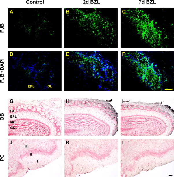
Figure 3.

Detection of degenerating neurons in OB and PC after bilateral zinc sulfate lesion. Neuronal degeneration was visualized by FJB staining (A–F, green) and amino cupric silver staining (G–L, black). FJB staining was increased 2 and 7 d after deafferentation in OB (B, C) compared with control (A), but not in PC (data not shown). Two and seven days after, degenerating neurons stained with silver were apparent in OB (H, I, respectively) but not in PC (K, L) compared with the control OB (G) and PC (J). Scale bars, 50 μm. EPL, External plexiform layer; GCL, granule cell layer; GL, glomerular layer; MCL, mitral cell layer.
