Figure 6.

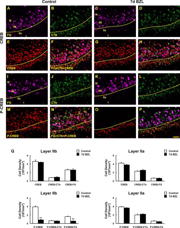
Phosphorylation of CREB in FG- and CTb-labeled pyramidal cells after deafferentation. A–E, G, I–M, O, FG- (violet; A, C, I, K) and CTb-labeled (green; B, D, J, L) pyramidal cells were colabeled with CREB (E, G) and phospho-CREB (p-CREB; M, O) antibody (red). F, H, Pyramidal cells traced by FG and CTb were both stained with CREB either in control (F) or at 7 d after bilateral zinc sulfate lesion (BZL) (H). N, P, Sections were also double labeled with phospho-CREB in control (N) and 7 d after BZL (P). The subset of pyramidal cells labeled with FG lost phospho-CREB labeling after BZL and most of these cells are located in layer IIb whereas CTb-labeled cells in layer IIa still express phospho-CREB even after zinc sulfate lesion (P). Q, Stereological cell counts revealed a significant decrease in phospho-CREB- and FG-double-labeled cell density after zinc sulfate lesion (**p < 0.01; ++p < 0.01). Data are means + SEM (n = 3). Scale bar, 50 μm.
