Figure 5.
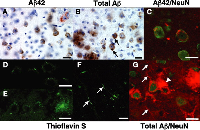
Intraneuronal Aβ accumulation in Tg6799: intracellular Aβ aggregates and plaque formation. Brain sections from representative 1.5- to 6-month-old Tg6799 mice were processed for immunocytochemistry with anti-Aβ antibodies (A–C, G) or stained with thioflavin S (D–F). Some sections were counterstained with hematoxylin (A, B) or with an antibody that recognizes the neuronal marker NeuN (green stain in C, G). Anti-Aβ antibodies used were specific for the C terminus of Aβ42 (A, brown; C, red) or recognized all forms of Aβ (B, 4G8, brown; G, R1282, red). Tg6799 ages were 1.5 (A, inset), 2 (A, C, D, F, G), 3 (E), and 6 (B) months. Intraneuronal Aβ accumulations were primarily observed as small puncta, although occasional large irregular accumulations were seen near the axon hillock (black arrows; B). Importantly, thioflavin S staining revealed intraneuronal Aβ aggregates consisting of β-pleated sheet amyloid accumulations within soma (punctate signals; D, E) and neurites (linear structures, white arrows; F). Note the thioflavin S-positive amyloid plaque (right; E). In some sections (G), an amyloid deposit (white arrowhead) was observed to originate from a neuron cell body with abnormal, disrupted-appearing morphology. This neuron had very little anti-NeuN antibody staining (green), indicating that it was likely in the process of degeneration. In addition, other neurons in the section appeared to be in various stages of degeneration (e.g., bottom right; G) and were also in close proximity to amyloid plaques (e.g., far left center; G). Note the presence of Aβ-filled neurites passing through the section (red linear structures, white arrows; G). Interestingly, plaque sizes were typically of the same magnitude as neuronal soma. Additional amyloid plaques can be seen at the bottom left in B and at the top right in G. For a movie of a three-dimensional reconstruction of the Z-stack sections in G, see supplemental material (available at www.jneurosci.org). Scale bars, 20 μm.

