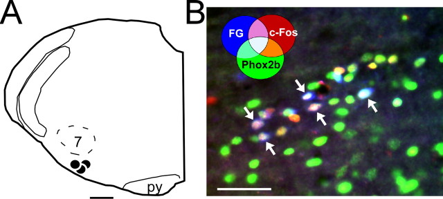
Figure 5.
Hypoxia-responsive NTS neurons with projections to RTN express Phox2b. A, NTS neurons with axonal projections to the RTN were prelabeled with the retrograde tracer FG in three rats. The FG injections (large black dots) were considered to be correctly placed because they were centered below the caudal and medial edge of the facial motor nucleus (7). py, Pyramidal tract. B, Awake rats were subjected to hypoxia and hypoxia-activated NTS neurons were identified by the presence of c-Fos-ir nuclei. The brain tissue was processed for simultaneous detection of FG (native blue fluorescence), Phox2b immunoreactivity (Alexa 488 fluorescence; green), c-Fos immunoreactivity (Cy3; red). The photomicrograph taken within the commissural portion of the NTS shows multiple aqua colored neurons (arrows) with RTN projections, activated by carotid body stimulation and expressing Phox2b. The color coding for other combinations of markers is indicated on the figure. Scale bars: A, 500 μm; B, 50 μm.

