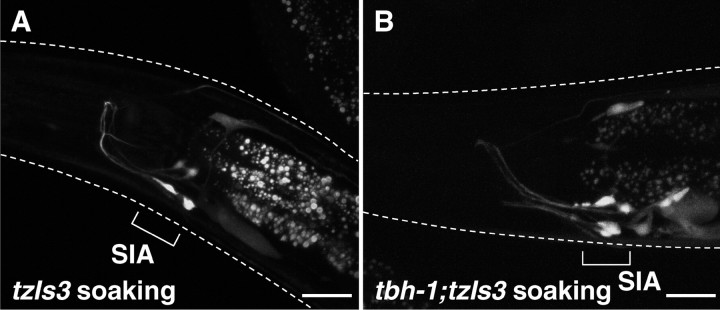
Figure 6.
Soaking-induced GFP expression in control animals and tbh-1 mutants carrying cre::gfp. Fluorescent images were taken from control animals (A) and tbh-1 mutants (B) carrying cre::gfp that were soaked in water in the presence of food. GFP expression in SIA neurons was observed in both control and tbh-1 mutants after soaking. White dotted lines indicate the outline of the heads of the animals. Scale bars, 20 μm.

