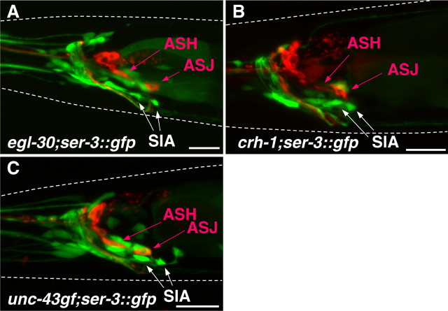
Figure 7.
SIA neurons in egl-30, crh-1, and unc-43 mutants. The fluorescent images were taken from egl-30(n686), crh-1(tz2), and unc-43(n498gf) mutants carrying ser-3::gfp. The expression of GFP and DiI staining of sensory neurons are shown in green and red, respectively. GFP expression was observed in the position of SIA neurons in egl-30 (A), crh-1 (B), and unc-43(n498gf) (C) mutants. White dotted lines indicate the outlines of the heads of the animals. Scale bars, 20 μm.

