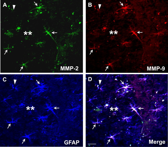
Figure 1.
MMP-2 and -9 immunoreactivity is selectively increased in activated astrocytes surrounding amyloid plaques in aged APP/PS1 mice. MMP-2 (A) or MMP-9 (B) immunoreactive cells (arrow) were prominent in cells surrounding Aβ deposits in the cortex or hippocampus of aged APP/PS1 mice. MMP-2 (A) and MMP-9 (B) immunoreactivity colocalized to GFAP-positive cells (C) showing morphological characteristics of reactive astrocytes (D, merged). Arrows indicate triple-labeled astrocytes with expression of MMP-2 and -9; arrowhead indicates a GFAP-positive astrocyte that is not immunoreactive to MMP-2 or -9; double asterisks indicate amyloid plaque. Scale bar, 10 μm.

