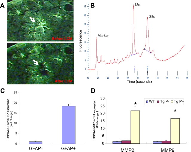
Figure 2.
MMP-2 and -9 mRNA levels are elevated in astrocytes surrounding amyloid plaques in aged APP/PS1 mice. A, Representative photomicrograph of GFAP-immunostained astrocytes in the cerebral cortex before and after laser capture microdissection. B, RNA electropherogram from microdissected astrocytes shows distinct 18S and 28S rRNA peaks indicating high RNA quality. C, Relative quantification of GFAP mRNA by real-time PCR revealed an 18-fold increase in GFAP-positive astrocytes in comparison with GFAP-negative neighboring cells, suggesting excellent enrichment of astrocyte-specific RNA. D, Real-time PCR quantification revealed significant increases in both MMP-2 and -9 in astrocytes surrounding amyloid plaques (Tg P+) compared with astrocytes distant from amyloid plaques (Tg P−) and astrocytes from control mice (WT). *p < 0.05; significant difference between wild-type and APP/PS1 astrocytes (n = 3). Error bars indicate mean ± SD.

