
Figure 2.
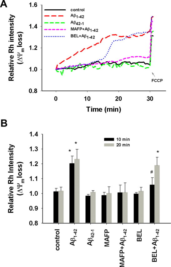
Aβ1–42 induced mitochondrial Δψm loss. Astrocytes were pretreated with MAFP (5 μm) or BEL (5 μm) for 30 min before exposure to Aβ1–42 (5 μm) or Aβ42–1 (5 μm) at t = 0. Rh 123 intensity was measured at every 30 s interval for 30 min, and then FCCP (0.5 μm) was added at the end of 30 min. A, Δψm loss in astrocytes attributable to Aβ1–42 but not Aβ42–1 was suppressed by MAFP, a specific inhibitor of both cPLA2 and iPLA2, whereas BEL, a specific inhibitor of iPLA2, only suppressed the Δψm loss for the initial ∼12 min. B, Quantitative relative Rh 123 intensities at the two representative time points 10 and 20 min. Aβ1–42 caused significant Δψm loss compared with control (*p < 0.02), whereas Aβ42–1, MAFP, or BEL alone had no effect on Δψm. MAFP completely suppressed the Δψm loss induced by Aβ1–42, whereas BEL only suppressed the Δψm loss at the initial 10 min after exposure to Aβ1–42 (#p > 0.05). Values are mean ± SD obtained from four independent experiments.