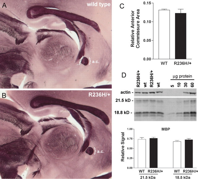
Figure 5.
Normal white matter architecture and MBP expression in the GFAP-mutant mice. A, B, Histochemical staining for myelin in the R236H/+ mice (B) shows normal anatomical structures when compared with wild-type mice (A) at 3 months of age. C, Area measures of the anterior commissure (a.c.) show no difference between wild-type (WT) and R236H/+ mice (n = 15 wild-type and 11 R236H/+ mice). D, Semiquantitative Western blotting was performed to compare MBP levels between R236H mutants and wild-type littermates at an earlier time point of P21. The first four lanes show examples of wild-type and R236H/+ protein (50 μg/lane); the last four lanes are representative of samples used to generate standard curves for actin versus MBP with the protein quantity indicated above the lane. No significant differences were found for 18.8 or 21.5 kDa isoforms (t test, n = 8 females at 21 d of age). Error bars indicate SD.

