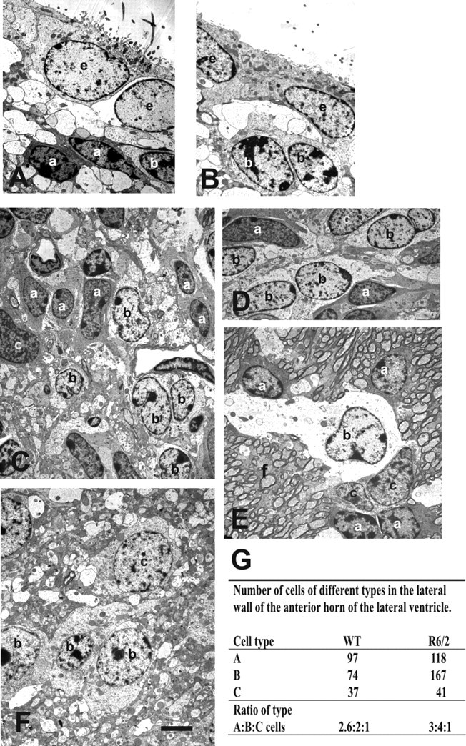
Figure 3.

Electron microscopy analysis show a specific increase number of B cells (b) in the subependyma of the anterior horn of the lateral ventricle of the R6/2 mice. A, B, Typical ependymal and subependymal cells from the WT anterior horn of the lateral ventricle. A (a) and B cells are seen among cell processes. C–F, Subependymal cells from the same region in R6/2 mice showing A, B, and C (c) cell types. This subependyma is thicker compared with the WT one in C. In D, a representative area with predominant B cells. In E, note the unusual location of the migratory A cells with B and C cells away from the lumen of the lateral ventricle, within the R6/2 striatal fibers (f). F, B and C cells were also farther away from the lumen of the ventricle than in WT mice, among cell processes. G shows the ratio of type A/B/C cells in the subependyma of the anterior horn of the lateral ventricle in WT and R6/2 mice. e, Ependymal cells. Scale bar: A, B, 1.8 μm; C, E, F, 1.6 μm.
