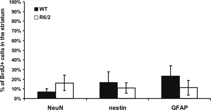
Figure 8.
Phenotypes of BrdU-positive cells in the striatum of WT and R6/2 mice. Specific markers were used to characterize BrdU+ cells 30 d after BrdU injection. There is a nonsignificant (t(5) = 1.188; p > 0.05) tendency for the R6/2 striatum to have a greater percentage of BrdU+ neurons (NeuN+ cells). The percentage of BrdU+ cells double labeled for GFAP in the R6/2 mice does not show a relative astrocytosis in the striatum compared with WT mice, but, given the greater number of BrdU+ cells, there are absolutely more astrocytes in the R6/2 striatum.

