
Figure 1.
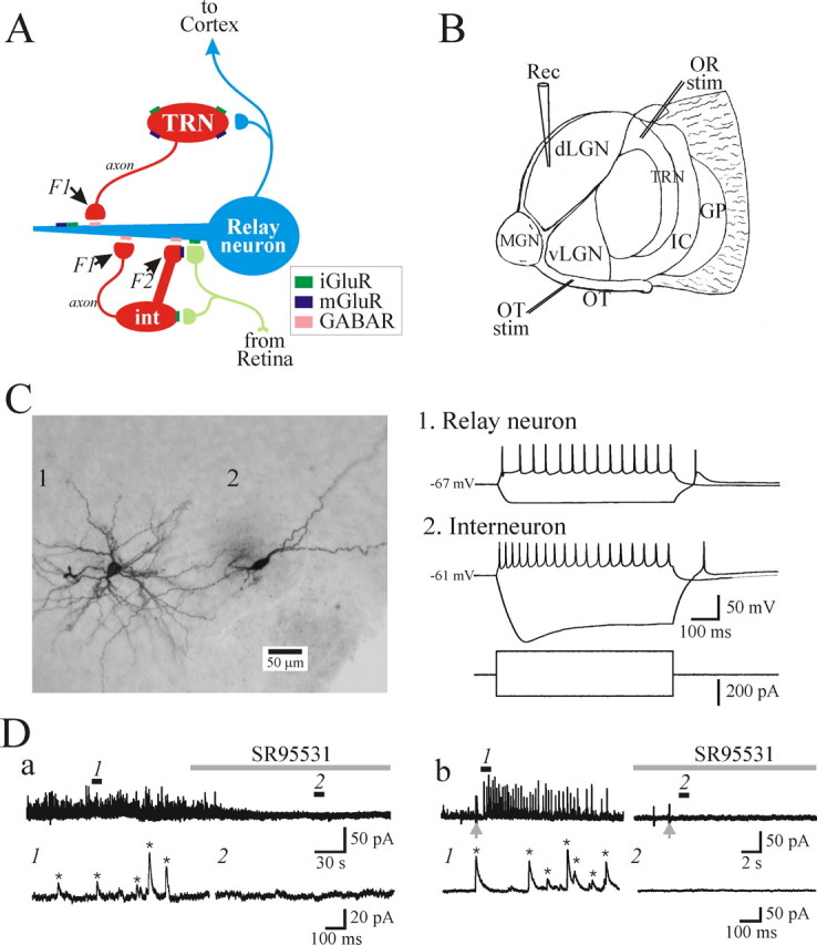
Characterization of GABAergic circuitry in the thalamus. A, Schematic diagram illustrating known inhibitory circuitry in the dLGN. Retinogeniculate axons form excitatory synapses onto relay neurons and dendrites of interneurons (int). These presynaptic dendrites of interneurons (F2 terminals) in turn form inhibitory synapses onto the relay neuron dendrites. Retinogeniculate axons also synapse near the interneuron cell body. Relay neurons also receive conventional axonal inputs (F1 terminals) from dLGN interneurons and TRN neurons. GABAR, GABA receptor. B, Schematic of the thalamic slice preparation used in the present study in which there are intact retinogeniculate and corticothalamic fibers. Retinal and cortical fibers were stimulated by placing the stimulation (stim) electrodes in optic tract (OT) and TRN, respectively. Rec, Recording electrode; OR, optic radiation; GP, globus pallidus; IC, internal capsule; MGN, medial geniculate nucleus; vLGN, ventral lateral geniculate nucleus. C, Representative cytoarchitecture (left) and electrophysiological responses (right) of an identified dLGN relay neuron (1) and interneuron (2). D, Current traces illustrating spontaneous (a) and optic tract-evoked (b) IPSCs recorded from dLGN relay neurons. The bottom traces are faster time bases referring to numbered regions above. The IPSC frequency and amplitude were quantified using Minianalysis software (Synaptosoft). The software detected events based on their amplitude, threshold, and decay time as indicated by the asterisk. Note that no events were detected in the presence of the GABAA receptor antagonist SR95531 (10 μm).