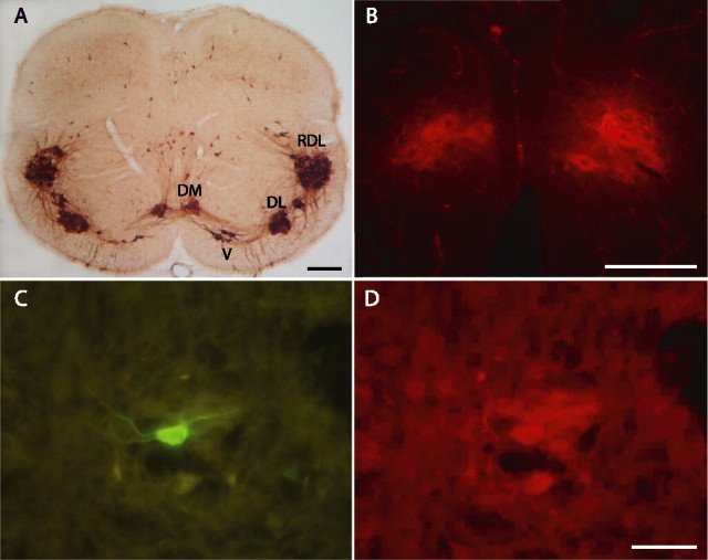
Figure 1.
Identification of pudendal motoneurons in neonate rats. A, Coronal section of the spinal cord from a 10-d-old rat showing the immunostaining of ChAT at segmental level L6. Pudendal motoneurons are grouped in DM and DL nuclei. The RDL, which projects to the hindleg, and the ventral nucleus (V), which projects to the pelvic diaphragm, are also detectable, as are isolated immunoreactive cells scattered in the medial and dorsal central gray. B, Photomicrograph illustrating the Fast DiI retrograde labeling of DM motoneurons. The spinal cord section is from a 10-d-old rat that had received an intraperitoneal injection of Fast DiI 24 h before. The animal was fixed by intracardiac perfusion of a solution containing 4% paraformaldehyde in 0.1 m PBS. Note, by comparing A and B, that Fast DiI-labeled DM nuclei are easily recognizable by their shape and position along the midline. C, D, Identification of a recorded biocytin-loaded DM motoneuron. C, Biocytin was visualized by treatment of the section with FITC-conjugated streptavidin. D, The biocytin-labeled motoneuron was immunoreactive for ChAT, detected with a Cy3-conjugated secondary antibody. Scale bars: A, 200 μm; B, D (for B–D), 100 μm.

