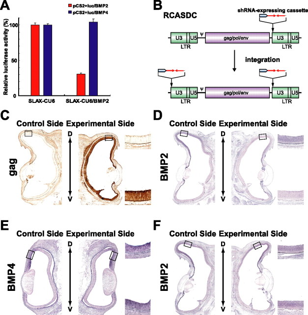
Figure 1.
The gene-specific knockdown system for the chick. A, Functional analysis of the vector-based shRNA in CEF cells. CEFs were transfected with a reporter plasmid (pCS2+luc/BMP2 or pCS2+luc/BMP4) and an shRNA expression plasmid (SLAX-CU6 or SLAX-CU6/BMP2). Luciferase activity was measured 48 h after transfection. SLAX-CU6/BMP2 selectively knocked down the luciferase expression from the reporter plasmid containing the BMP2 sequence. Luciferase activity in the cells transfected with an empty vector SLAX-CU6 was set to 100%. Error bars indicate SD. B, Schematic representation of the RCASDC retroviral vector. The shRNA-expressing cassette was inserted into the U3 region of the 3′ LTR of the RCASDC. The resultant provirus acquires the shRNA-expressing cassette sequences at both ends by duplication of the LTR during the reverse transcription and integrates them into the genome of transfected cells. C, Expression of viral gag protein in the right eye transfected. RCASDC-CU6/BMP2 was introduced to the embryo at E1.5 by electroporation. Viral gag protein was visualized at E8 by immunostaining with monoclonal antibody 3C2. D, E, Coronal section in situ hybridization of control retinas (left) and RCASDC-CU6/BMP2-electroporated retinas (right). Expression of BMP2 at E8 was markedly reduced in the retina by electroporation of RCASDC-CU6/BMP2 (D). Expression of BMP4 at E4 was not altered by electroporation of RCASDC-CU6/BMP2 (E). F, Coronal section in situ hybridization of the retinas at E8 electroporated with RCASDC-CU6/EGFP (right) at E1.5. Expression of BMP2 was not changed by the expression of shRNA for EGFP. The boxed regions in the control retina (left) and manipulated retina (right) are enlarged on the top right and bottom right sides, respectively (C–F).

