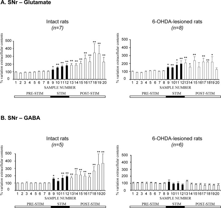
Figure 3.
Extracellular glutamate (A) and GABA (B) levels, determined at 15 min intervals, in the SNr ipsilateral to the stimulation in intact and 6-OHDA-lesioned rats under basal conditions and during 1 h of STN–HFS with an intensity inducing forelimb dyskinesia (I1, >60 μA). The prestimulation period (PRE-STIM) corresponds to fractions 1–8 of the dialysates; the stimulation period (STIM), indicated by the black horizontal bar, corresponds to fractions 9–12 (4 fractions) and the poststimulation period (POST-STIM) corresponds to fractions 13–20 (8 fractions). The mean ± SEM of the eight PRE-STIM dialysates, collected before STN–HFS, was used to determine baseline levels. Results are expressed as a percentage of variation of this baseline value. Each percentage corresponds to the mean ± SEM variations calculated for five to eight animals. Note that Glu levels increased in both intact and 6-OHDA-lesioned rats, whereas GABA levels increased only in intact animals. *p < 0.05, **p < 0.01 versus baseline values. Error bars correspond to the SEM.

