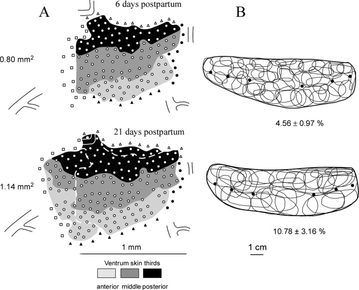
Figure 3.
Cortical map expansion and RF sharpening induced by nursing. A, Two somatotopic maps were derived from the same rat at days 6 and 21 after nursing initiation. These ventrum skin representations are composed of territories in which RFs were centered on the anterior, middle, and posterior thirds of the ventrum skin. Open circles indicate recording sites in which cutaneous responses were recorded. Open squares mark cortical sites displaying noncutaneous responses elicited by strong pressure on skin. Filled and open triangles refer to sites excited by forelimb and hindlimb stimulation, respectively. The broken lines on the second map outline the main cortical zones over which representational expansion occurred. Constant vascular landmarks are shown in each map to facilitate comparison. B, RFs defined on the ventrum skin at the recording sites shown in the corresponding maps. The heavy dark lines outline the ventrum skin. Filled circles indicate the location of nipples. For sake of clarity, ∼30% of the recorded RFs are not shown. Mean ± SD values of map areal extent and normalized (with reference to total ventrum skin area) RF sizes are indicated for each mapping session. Note that, at day 21 postpartum, when daily nursing time was reduced to ∼30%, the ventrum map was still in expansion, whereas the sizes of neuron RFs within the corresponding cortical area were as large as those recorded before nursing.

