Figure 6.

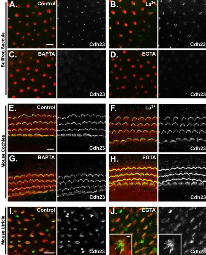
Cdh23 immunolocalization in hair cells after loss of tip links. Each panel depicts a single confocal section of whole mounted bullfrog saccule, mouse cochlea, or mouse utricle. The left panel for each group shows the merged image of actin (red) and Cdh23 immunolocalization (green). The right panel depicts the Cdh23 immunolabeling channel alone. A, Cdh23 immunolocalization in the bundles of bullfrog saccular hair cells. B–D, Cdh23 immunolocalization in the bundles of bullfrog saccular hair cells after treatment with 5 mm La3+, BAPTA, or EGTA, respectively. E, Cdh23 immunolocalization in the bundles of mouse cochlear hair cells (P1 plus 1 d in vitro). F–H, Cdh23 immunolocalization in the bundles of mouse cochlear hair cells after treatment with 5 mm La3+, BAPTA, or EGTA, respectively. I, Cdh23 immunolocalization in the bundles of mouse (P6) utricular hair cells. Asterisks demark representative small hair bundles, which are intensely labeled. An arrowhead denotes an example of stereociliary tip labeling. J, Cdh23 immunolocalization in the bundles of mouse utricular hair cells (P6) after treatment with 5 mm EGTA. Inset, A small hair bundle with labeling along the length of the stereocilium. This example is from a P3 mouse utricule treated with EGTA at 4°C. Similar results were seen with P6 utricles treated at room temperature. Scale bars: A, E, I, (for A–D, E–H, I–J), 10μm; J, inset, 2 μm.
