
Figure 7.
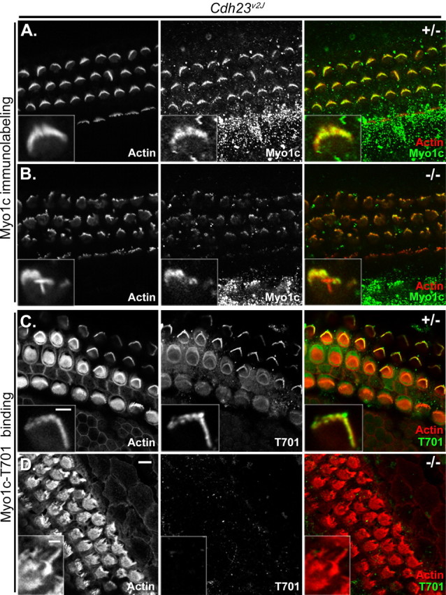
Myo1c and Myo1c receptor localization in cochlear hair cells of Cdh23v2J mice. Each panel shows three rows of outer and one row of inner hair cells of heterozygous (+/−) or homozygous (−/−) Cdh23v2J mutant mice (P2–P4). The left column of each panel shows phalloidin labeling alone, and the middle column of each panel shows the bound Myo1c antibody or Myo1c-T701 binding alone. The right column of each panel shows the overlay of phalloidin-labeled filamentous actin (red) and the binding of either a Myo1c antibody (green; A, B) or the Myo1c-T701 fragment (green; C, D). A–D, Cochlear cultures from Cdh23v2J mice. A, B, Endogenous Myo1c is present in stereocilia of both the Cdh23v2J heterozygous and homozygous mice. C, The Myo1c receptor is detected in the stereocilia of the Cdh23v2J heterozygous mice. D, Myo1c receptor is undetectable in the stereocilia of Cdh23v2J homozygous mice. Insets depict the hair bundles of outer hair cells. Scale bar: (in D) 5 μm; (in C, inset), A, B, C, insets, 2 μm; D, inset, 2 μm.