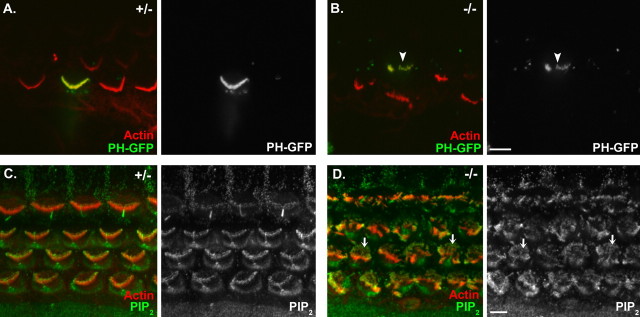
Figure 9.
PIP2 is present in the stereocilia of Cdh23v2J mice. A, B, Localization of PIP2 in a cochlear apical coil from a heterozygous (A) and a homozygous (B) Cdh23v2J mouse using the PH domain of PLCδ1 conjugated to EGFP as a fluorescent indicator of PIP2 localization. C, D, Immunolocalization of PIP2 in a cochlear basal coil from a heterozygous (C) and a homozygous (D) Cdh23v2J mouse. All images are single confocal sections. In the merged image to the left, phalloidin-labeled filamentous actin is depicted in red, and PLCδ1PH-EGFP or an anti-PIP2 antibody is green. In the image on the right, PLCδ1PH-EGFP or the antibody labeling alone is shown. The arrowhead in B denotes a fluorescent mutant hair bundle. The arrows in D denote microvilli labeling. Scale bars: (in B) A, B, 5 μm; (in D) C, D, 5 μm.

