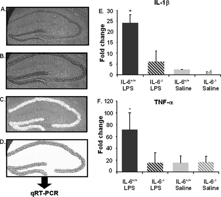
Figure 5.
A–D, Laser capture microdissection of the hippocampal neuronal layer. A–D, Stained section (A), captured neuronal layer (B), section with removed hippocampus (C), and isolated hippocampal neuronal layer (D). E, F, Percent fold change for mRNA cytokine levels for IL-6+/+ (solid filled bars) and IL-6−/− (striped bars) mice that received a single intraperitoneal injection of LPS (black-filled bars) or saline (gray-filled bars) 4 h before kill. IL-1β mRNA (E) and TNF-α (F). Data are presented as mean±SEM. nd, Cytokine levels were nondetectable. *Significant difference between groups as determined by Fisher's protected least-significant difference (p < 0.05).

