Figure 6.

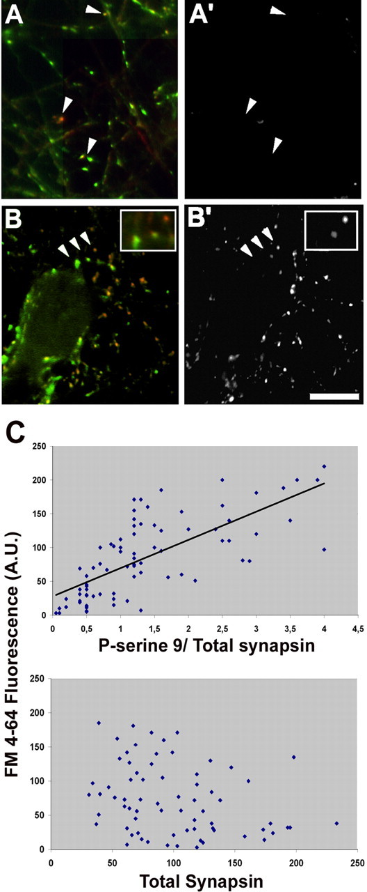
Depolarization-induced SV recycling correlates with the intensity of synapsin phosphorylation at site 1. A–B′, Rat hippocampal neurons were loaded for 1 min with FM4-64 (A′, B′) in either KRH (A′) or depolarizing solution (B′), fixed, and processed for retrospective double immunofluorescence (A, B) with an antibody that recognizes all synapsin isoforms (green) and with a phosphosite-1-specific anti-synapsin antibody (red). Arrowheads in A and A′ point to unstimulated synapses showing a high level of site-1 phosphorylation not accompanied by detectable FM4-64 internalization. Arrowheads in B and B′ point to stimulated synapses (shown at higher magnification in the insets) in which the intensity of staining for site 1-phosphorylated synapsin correlates with the intensity of FM4-64 labeling. Scale bar, 10 μm. C, Quantitative correlation between apparent stoichiometry of synapsin phosphorylation at site 1 (top) (r = 0.71; p < 0.001, Pearson's test) or total synapsin immunoreactivity (bottom) (r = −0.28; p > 0.1) and the level of FM4-64 uptake induced by depolarization. Each point corresponds to one synapse. The intensity of the labeling for phosphorylated synapsin is normalized for the total synapsin immunoreactivity in that terminal. A.U., Arbitrary units.
