Figure 1.
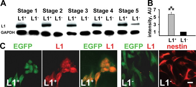
Transfected ES cells express L1 at all stages of differentiation. A, Immunoblot analysis of L1 expression in transfected (L1 +) and mock-transfected (L1 −) GFP + ES cells differentiated by the five-stage differentiation protocol at day 2 of stages 1 and 2, day 6 of stages 3 and 4, and day 7 of stage 5. Note that transgenic L1 is expressed throughout all stages of differentiation. Immunoblot analysis of glyceraldehyde 3-phosphate dehydrogenase (GAPDH) is shown as a loading control. B, Quantification of the intensity in the immunoblot analysis of L1 expression in stage 5. Arbitrary units (AU) normalized for L1 expression in L1 − cells are shown (mean ± SEM). Student's t test was performed for statistical analysis (***p < 0.001). C, L1 overexpression at the cell surface was evaluated using anti-L1 (red) immunostaining and fluorescence light microscopy of live L1 − and L1 + cells at stage 4. Note the expression of GFP (green) in L1 − and L1 + cells. Neural differentiation of ES cells was confirmed by immunostaining of cells differentiated to stage 4 for nestin (red). Scale bar, 10 μm. EGFP, Enhanced GFP.

