
Figure 2.
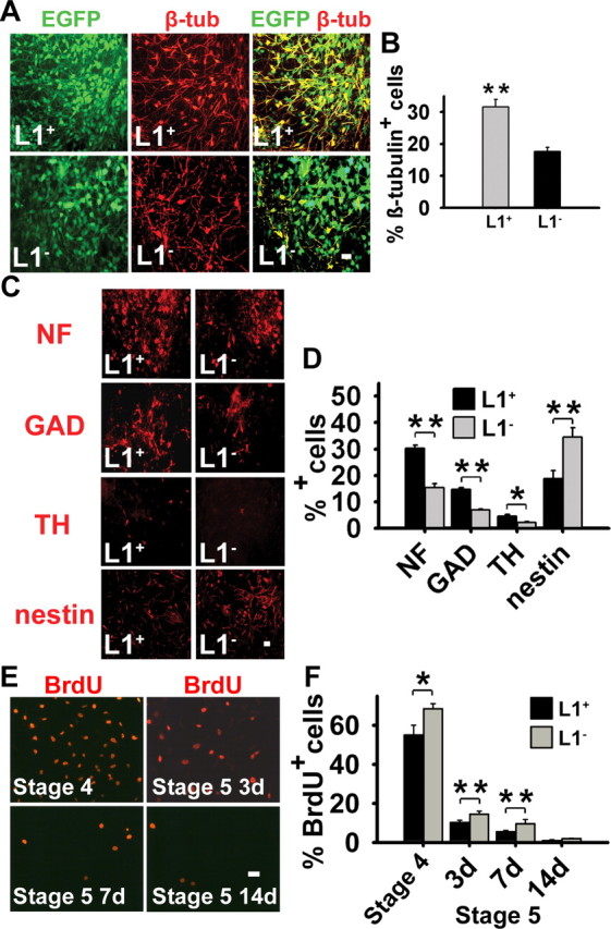
L1 expression increases neuronal differentiation and decreases proliferation in vitro. A, Generation of neurons from L1 − and L1 + ES cells was determined at day 7 of stage 5 by immunostaining for β-tubulinIII (β-tub; red). GFP + cells, green; β-tubulinIII/GFP + cells, yellow. The proportion of β-tubulinIII + cells of all GFP + cells was greater in L1 + cells than in L1 − cells. Scale bar, 20 μm. EGFP, Enhanced GFP. B, Percentages of β-tubulinIII + cells of all GFP + cells are shown at day 7 of stage 5 (mean ± SEM). Student's t test was performed for statistical analysis (**p < 0.01). C, At day 14 of stage 5, mature neuronal proteins NF-200 (NF), GAD, and TH and the neural stem cell protein nestin (all in red) were determined by immunocytochemistry in L1 − and L1 + cells. The proportion of NF +, GAD +, and TH + cells was greater in the L1 + than in the L1 − cells, whereas the proportion of nestin + cells was higher in L1 − cells. Scale bar, 10 μm. D, Percentages of NF +, GAD +, TH +, and nestin + cells at day 14 of stage 5 (mean ± SEM). *p < 0.05, **p < 0.01 (Student's t test). E, BrdU incorporation was determined at day 7 of stage 4 and at days 3, 7, and 14 of stage 5 by immunostaining for BrdU (red) after a pulse labeling of 8 h with 10 mm BrdU. Scale bar, 10 μm. F, Percentages of BrdU + cells of all cells are shown at day 7 of stage 4 and days 3, 7, and 14 of stage 5 (mean ± SEM). Student's t test was performed for statistical analysis (*p < 0.05; **p < 0.01). 3d, Day 3; 7d, day 7; 14d, day 14.