
Figure 4.
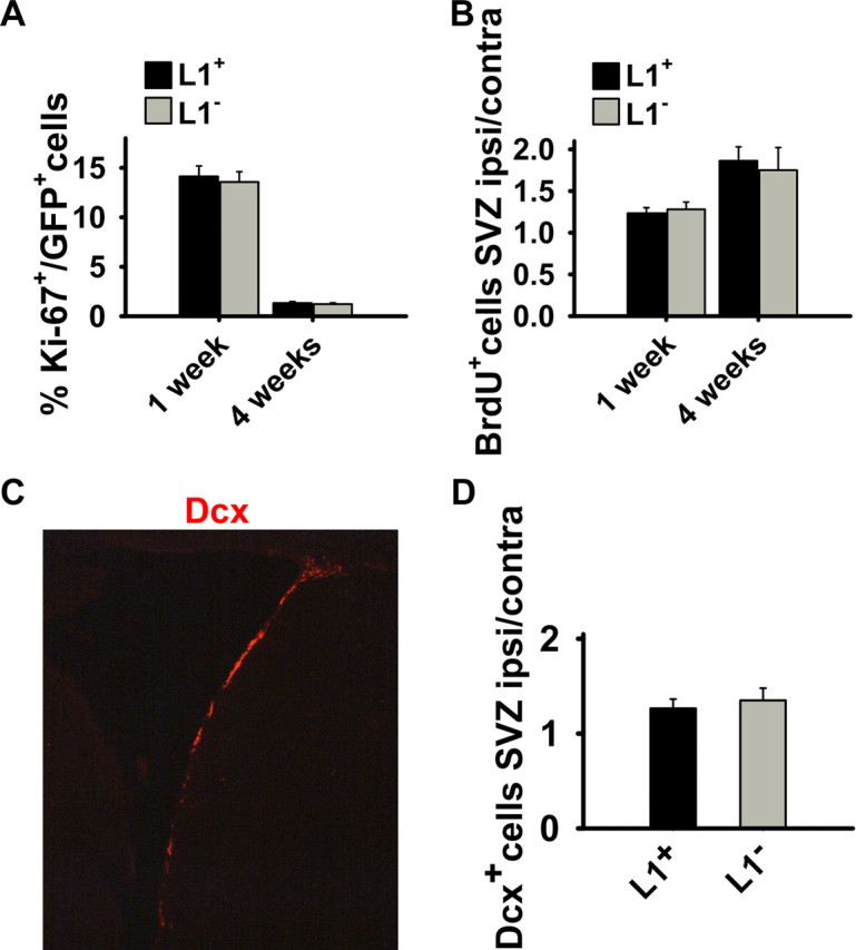
Analysis of proliferation in grafted cells by Ki-67 immunolabeling and of neurogenesis in the SVZ by BrdU incorporation and expression of DCX 1 and 4 weeks after transplantation. A, The fraction of Ki-67 + cells of all GFP + transplanted cells is given 1 and 4 weeks after transplantation of L1 + (n = 4) and L1 − (n = 4) cells. No difference was observed. B, Mice transplanted with L1 + (n = 4) or L1 − (n = 4) cells into the lesioned striatum were labeled with BrdU on days 3–7 after transplantation and killed 7 d and 4 weeks after grafting. The ratio of BrdU + cells in the SVZ ipsilateral and contralateral to the lesioned hemisphere is shown 1 and 4 weeks after transplantation. No difference was observed between the L1 + and L1 − groups. C, Immunohistochemical staining for DCX (red) in the SVZ ipsilateral to the lesion in sham-injected animals 4 weeks after grafting. D, Immunostaining for DCX in the SVZ was assessed 4 weeks after grafting. The ratio of DCX + cells in the SVZ ipsilateral and contralateral to the lesioned and transplanted hemisphere is given for mice transplanted with L1 + (n = 4) or L1 − (n = 4) cells into the lesioned striatum. No difference between L1 + and L1 − groups was observed. Error bars indicate SEM. ipsi, Ipsilateral; contra, contralateral.