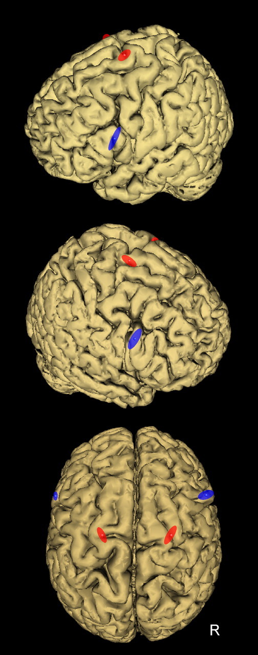
Figure 2.

Location of the TMS coil positions to induce virtual lesion of PMv (blue) and PMd (red). To stimulate PMv, the coil was positioned over the caudal portion of the pars opercularis of the inferior frontal gyrus (BA44), corresponding to the following normalized MNI coordinates: −60 ± 2, 16 ± 3, 23 ± 9 mm (x, y, z, mean ± SD; n = 10) and 56 ± 6, 16 ± 4, 26 ± 9 mm (n = 6) for the left and right PMv, respectively. To target PMd, the coil was positioned over the superior portion of the precentral gyrus, as delimited by the superior frontal sulcus. The mean MNI coordinates of stimulation sites for the left and right PMd were, respectively, −22 ± 3, −4 ± 4, 71 ± 4 mm (x, y, z, mean ± SD; n = 10) and 24 ± 4, −5 ± 6, 72 ± 3 mm (n = 6). Each ellipse was centered on the mean MNI coordinates of PMv and PMd stimulation points, and their surface shows the 95% confidence interval of the normalized coordinates calculated for each subject.
