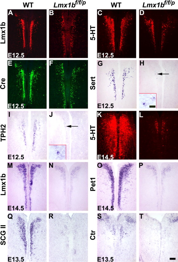
Figure 3.

Downregulation and/or loss of molecular markers in the rostral part of the hindbrain of Lmx1bf/f/p mice compared with wild-type mice. A–F, Immunocytochemical staining showed that Lmx1b (A, B), 5-HT (C, D), and Cre (E, F) expression was downregulated in Lmx1bf/f/p mice (B, D, F) compared with the control (A, C, E) at E12.5. G–J, Expression of Sert and TPH2 detected by in situ hybridization was virtually lost in Lmx1bf/f/p mice (H, J) at E12.5. Arrows indicate a few remaining Sert+ and TPH2+ cells. Small insets in H and J showed higher magnification of Sert+ and TPH2+ cells. K, L, 5-HT staining was almost lost in Lmx1bf/f/p mice (L) by E14.5. M–P, Similar downregulation of Lmx1b and Pet1 in Lmx1bf/f/p mice (N, P) compared with wild-type mice (M, O) at E14.5. Q–T, Expression of SCGII and Ctr was almost lost in Lmx1bf/f/p mice (R, T) at E13.5. WT, Wild type. Scale bar: 100 μm; inset, 20 μm.
