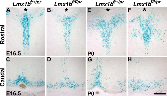
Figure 4.
Examination of central 5-HT neurons by X-gal staining. A–D, X-gal staining of 5-HT neurons in the rostral (A, B) and caudal (C, D) part of the hindbrain of Lmx1bf/+/pr (A, C) and Lmx1bf/f/pr (B, D) mice at E16.5. X-gal staining patterns were comparable between Lmx1bf/+/pr and Lmx1bf/f/pr mice. E–H, X-gal staining of 5-HT neurons in the rostral (E, F) and caudal (G, H) part of the hindbrain of Lmx1bf/+/pr (E, G) and Lmx1bf/f/pr (F, H) mice at P0. X-gal staining pattern was also similar in Lmx1bf/f/pr mice compared with Lmx1bf/+/pr mice. Asterisk (*) indicates the cerebral aqueduct. Scale bar, 100 μm.

