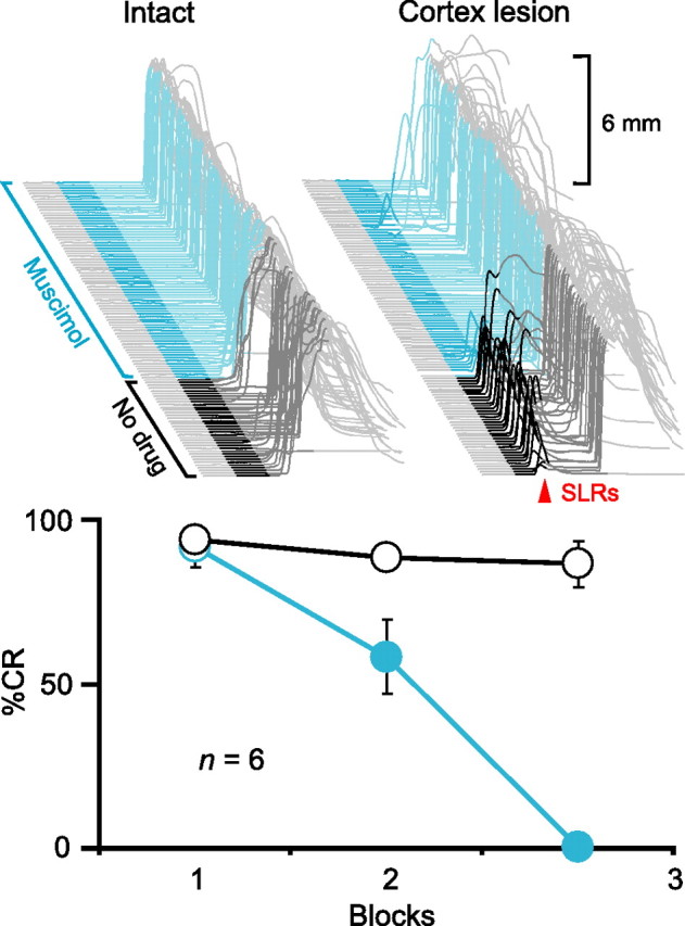
Figure 2.

Expression of CRs and SLRs requires activity in the AIN. Top left, Sample traces from an animal during a test session in which the GABAA agonist muscimol infused into the AIN abolished CRs. Top right, Sample traces from the same animal in which muscimol abolished SLRs (red arrowhead) unmasked by electrolytic lesions of the cerebellar cortex. In this and all subsequent figures, sample eyelid traces are arranged chronologically from front to back with the time of CS presentation highlighted (dark gray, no drug; color, after start of infusion) and the first 200 ms of the CS darkened to emphasize SLRs (upward deflection, eyelid closure). Scale bars denote the magnitude of full eyelid closure (6 mm). Bottom, Summary data showing percentage CRs as a function of 36-trial blocks during test sessions with muscimol (cyan) or a standard training session (black and white) before the cerebellar cortex lesion. For summary data in this and all subsequent figures, circles and bars (open, no drug; filled, after start of infusion) indicate CRs and SLRs, respectively. Error bars show SEM.
