
Figure 1.
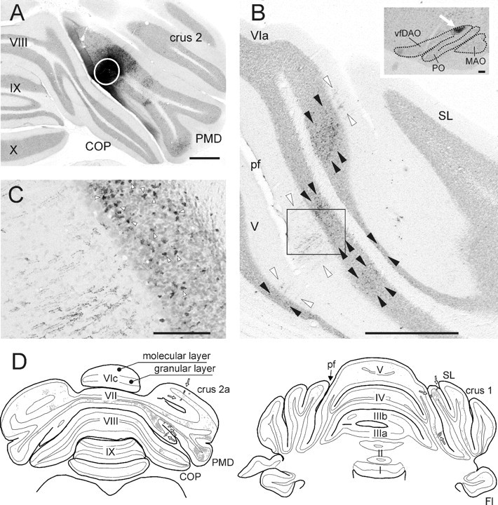
Photomicrographs showing aspects of the CTb injection site and resultant labeling of climbing and mossy fiber collaterals in case AP03 together with a selection of plots of transverse sections through cerebellum of case AP09. A, Photomicrograph of the CTb injection site, indicated by a white circle, in the paramedian lobule. The injection was mostly restricted to the molecular and granular layers of the electrophysiologically identified C1 zone, which was verified by the presence of retrograde labeled cells in the contralateral vfDAO (B, inset). B, Photomicrograph showing part of a section of the SL and anterior lobe of the same case. Note the characteristic appearance of CTb-labeled climbing fibers in the molecular layer (between white arrowheads) and the numerous labeled mossy fiber rosettes within the granular layer (between black arrowheads). Also note that the regions with climbing fiber terminals are aligned in the mediolateral plane but that the regions with mossy fiber terminal rosettes are also found at other mediolateral positions. The inset shows retrogradely labeled cells in the dorsal accessory olive (arrow) of the contralateral inferior olive. C, Magnification of the boxed rectangle in B showing the boundary of granular and molecular layer. The arrowheads indicate some of the labeled mossy fiber rosettes that are located directly adjacent to labeled climbing fiber terminal arborizations. D, Two plots of transverse sections through the posterior and anterior cerebellum, respectively, and indicating the distribution of CTb labeling resulting from a PMD injection in the electrophysiologically identified C2 zone (case AP09). Labeled mossy fiber rosettes (small gray dots: one dot equals one rosette) and climbing fiber terminals (black dots indicated by arrows) are indicated. Scale bars: A, B, 1 mm; B, inset, 100 μm; C, 100 μm. I–X, Lobules I–X; Fl, flocculus; pf, primary fissure.