Figure 12.
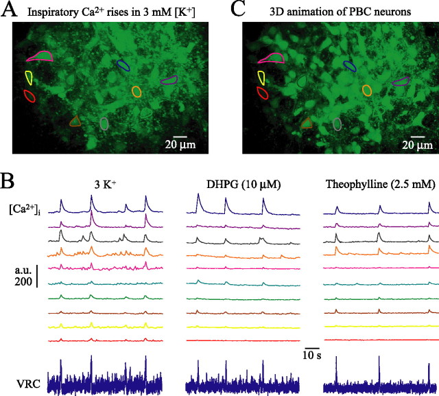
Confocal imaging of activity and morphology of inspiratory PBC neurons. A, Fluo-4-AM-stained PBC neurons outlined by regions of interest at −0.59 in a mPBC[500/−0.64] W-P1.5 slice. The supplemental movie (available at www.jneurosci.org as supplemental material) shows 90 s recording in 3 mm [K+] of rhythmic Ca2+ oscillations in these neurons that are in phase with inspiratory population activity recorded from the contralateral PBC (B, bottom left trace). B, Fluo-4-AM fluorescence intensity is plotted in arbitrary units (a.u.) against time. After washout of rhythm in 3 mm [K+], PBC bursting and Ca2+ oscillations were reactivated by DHPG and theophylline. C, 3D animation (supplemental material, available at www.jneurosci.org) showing gross morphology of PBC neurons and neighboring nonrhythmic cells obtained from z-stack image series (0.5 μm single step) encompassing areas starting 7.5 μm above to 7.5 μm below image plane of A.

