
Figure 3.
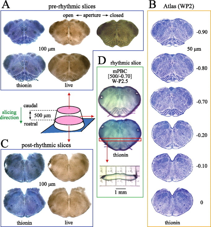
On-line histology for generating calibrated PBC slices. The diagram shows newborn rat brainstem glued with rostral side to metal plate attached to vibratome stage. Serial sectioning in caudorostral direction provides 100-μm-thin prerhythmic sections (A), which are compared with a W-P2 rat atlas (B) (supplemental material, available at www.jneurosci.org) to determine cutting level and caudal boundary of the rhythmic slice, in this case −0.70 mm relative to VIIc. As shown for the second-to-last prerhythmic slice (top row), resolution can be enhanced by adjusting the condensor aperture or focus. Sections are photographed, fixed for thionin staining, and rephotographed for higher resolution off-line analysis. C, Postrhythmic slices taken after cutting the rhythmic slice are used for on- and off-line analysis of rostral slice boundary. D, Fixed rhythmic slice stained with thionin to define caudal (top image) and rostral (bottom image) surfaces. The thickness of the rhythmic slice, here 500 μm, is determined by sectioning the horizontal strip at level of VRC/PBC. Combined on-line/off-line analysis revealed caudal and rostral slice boundaries at −0.70 and −0.20, respectively, and that the PBC is close to the middle of the slice (Fig. 2A). Sections in A, C, and D are from one individual WP2.5 brainstem. Thus, this calibrated PBC slice is described as “mPBC[500/−0.70]W-P2.5.”